

Which Of The Following Does The Autonomic Nervous System Most Directly Control?
The Autonomic Nervous System
Divisions of the Autonomic Nervous System
OpenStaxCollege
Learning Objectives
Past the stop of this department, you will be able to:
- Proper name the components that generate the sympathetic and parasympathetic responses of the autonomic nervous system
- Explicate the differences in output connections within the two divisions of the autonomic nervous organisation
- Describe the signaling molecules and receptor proteins involved in communication within the two divisions of the autonomic nervous system
The nervous system can be divided into ii functional parts: the somatic nervous system and the autonomic nervous organization. The major differences between the two systems are evident in the responses that each produces. The somatic nervous organisation causes wrinkle of skeletal muscles. The autonomic nervous system controls cardiac and smooth muscle, as well as glandular tissue. The somatic nervous system is associated with voluntary responses (though many can happen without conscious awareness, similar breathing), and the autonomic nervous organization is associated with involuntary responses, such as those related to homeostasis.
The autonomic nervous organization regulates many of the internal organs through a balance of 2 aspects, or divisions. In addition to the endocrine system, the autonomic nervous organization is instrumental in homeostatic mechanisms in the body. The two divisions of the autonomic nervous organization are the sympathetic division and the parasympathetic sectionalisation. The sympathetic system is associated with the fight-or-flying response, and parasympathetic activeness is referred to by the epithet of rest and digest. Homeostasis is the balance between the two systems. At each target effector, dual innervation determines activity. For instance, the heart receives connections from both the sympathetic and parasympathetic divisions. Ane causes heart rate to increase, whereas the other causes heart rate to decrease.

Watch this video to learn more about adrenaline and the fight-or-flight response. When someone is said to accept a rush of adrenaline, the paradigm of bungee jumpers or skydivers normally comes to mind. But adrenaline, also known as epinephrine, is an important chemical in coordinating the body's fight-or-flight response. In this video, you look inside the physiology of the fight-or-flight response, as envisioned for a firefighter. His torso's reaction is the result of the sympathetic division of the autonomic nervous system causing system-broad changes equally it prepares for extreme responses. What two changes does adrenaline bring well-nigh to help the skeletal muscle response?
Sympathetic Segmentation of the Autonomic Nervous System
To respond to a threat—to fight or to run abroad—the sympathetic organisation causes divergent furnishings as many different effector organs are activated together for a common purpose. More oxygen needs to be inhaled and delivered to skeletal muscle. The respiratory, cardiovascular, and musculoskeletal systems are all activated together. Additionally, sweating keeps the backlog heat that comes from muscle contraction from causing the body to overheat. The digestive system shuts downwards so that blood is not absorbing nutrients when information technology should be delivering oxygen to skeletal muscles. To coordinate all these responses, the connections in the sympathetic organisation diverge from a limited region of the central nervous system (CNS) to a broad assortment of ganglia that project to the many effector organs simultaneously. The complex set of structures that compose the output of the sympathetic organization make it possible for these disparate effectors to come together in a coordinated, systemic change.
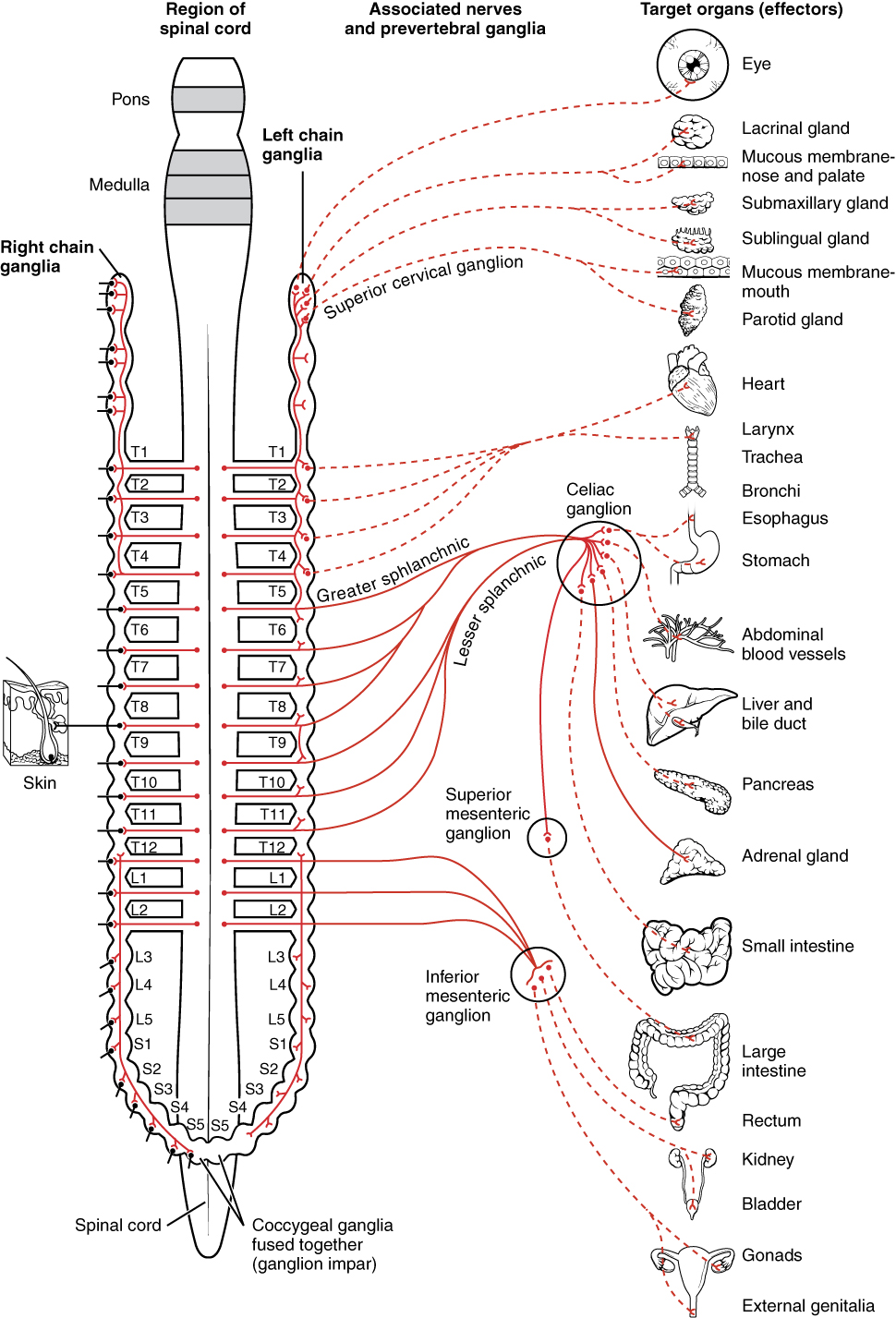
The sympathetic partitioning of the autonomic nervous system influences the various organ systems of the trunk through connections emerging from the thoracic and upper lumbar spinal cord. It is referred to as the thoracolumbar system to reflect this anatomical ground. A primal neuron in the lateral horn of any of these spinal regions projects to ganglia adjacent to the vertebral cavalcade through the ventral spinal roots. The majority of ganglia of the sympathetic organisation belong to a network of sympathetic chain ganglia that runs alongside the vertebral cavalcade. The ganglia appear equally a series of clusters of neurons linked by axonal bridges. In that location are typically 23 ganglia in the chain on either side of the spinal cavalcade. Iii correspond to the cervical region, 12 are in the thoracic region, four are in the lumbar region, and four represent to the sacral region. The cervical and sacral levels are not continued to the spinal cord directly through the spinal roots, just through ascending or descending connections through the bridges within the concatenation.
A diagram that shows the connections of the sympathetic system is somewhat similar a excursion diagram that shows the electrical connections between different receptacles and devices. In [link], the "circuits" of the sympathetic arrangement are intentionally simplified.
Connections of Sympathetic Division of the Autonomic Nervous Organization
Neurons from the lateral horn of the spinal cord (preganglionic nerve fibers – solid lines)) projection to the chain ganglia on either side of the vertebral column or to collateral (prevertebral) ganglia that are anterior to the vertebral column in the abdominal cavity. Axons from these ganglionic neurons (postganglionic nerve fibers – dotted lines) and then projection to target effectors throughout the body.
To continue with the analogy of the circuit diagram, there are 3 different types of "junctions" that operate within the sympathetic system ([link]). The first type is nigh direct: the sympathetic nerve projects to the concatenation ganglion at the same level as the target effector (the organ, tissue, or gland to exist innervated). An instance of this type is spinal nervus T1 that synapses with the T1 chain ganglion to innervate the trachea. The fibers of this branch are called white rami communicantes (singular = ramus communicans); they are myelinated and therefore referred to as white (meet [link]a). The axon from the fundamental neuron (the preganglionic fiber shown every bit a solid line) synapses with the ganglionic neuron (with the postganglionic fiber shown every bit a dashed line). This neuron then projects to a target effector—in this instance, the trachea—via gray rami communicantes, which are unmyelinated axons.
In some cases, the target effectors are located superior or junior to the spinal segment at which the preganglionic cobweb emerges. With respect to the "wiring" involved, the synapse with the ganglionic neuron occurs at chain ganglia superior or junior to the location of the key neuron. An case of this is spinal nerve T1 that innervates the middle. The spinal nerve tracks up through the chain until it reaches the superior cervical ganglion, where it synapses with the postganglionic neuron (see [link]b). The cervical ganglia are referred to as paravertebral ganglia, given their location adjacent to prevertebral ganglia in the sympathetic chain.
Non all axons from the central neurons terminate in the chain ganglia. Additional branches from the ventral nervus root continue through the concatenation and on to i of the collateral ganglia as the greater splanchnic nerve or lesser splanchnic nervus. For example, the greater splanchnic nerve at the level of T5 synapses with a collateral ganglion outside the chain before making the connection to the postganglionic nerves that innervate the stomach (see [link]c).
Collateral ganglia, as well called prevertebral ganglia, are situated anterior to the vertebral column and receive inputs from splanchnic nerves equally well as cardinal sympathetic neurons. They are associated with decision-making organs in the abdominal cavity, and are also considered part of the enteric nervous system. The 3 collateral ganglia are the celiac ganglion, the superior mesenteric ganglion, and the inferior mesenteric ganglion (encounter [link]). The word celiac is derived from the Latin word "coelom," which refers to a body cavity (in this instance, the abdominal cavity), and the word mesenteric refers to the digestive system.
Sympathetic Connections and Chain Ganglia
The axon from a cardinal sympathetic neuron in the spinal string tin can project to the periphery in a number of different ways. (a) The cobweb can projection out to the ganglion at the same level and synapse on a ganglionic neuron. (b) A branch can project to more superior or inferior ganglion in the chain. (c) A co-operative can project through the white ramus communicans, just not terminate on a ganglionic neuron in the chain. Instead, it projects through one of the splanchnic nerves to a collateral ganglion or the adrenal medulla (non pictured).
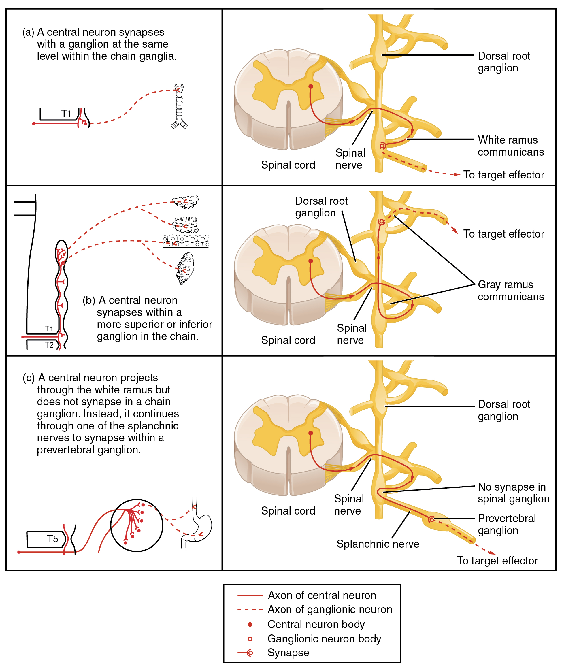
An axon from the central neuron that projects to a sympathetic ganglion is referred to every bit a preganglionic fiber or neuron, and represents the output from the CNS to the ganglion. Because the sympathetic ganglia are adjacent to the vertebral column, preganglionic sympathetic fibers are relatively short, and they are myelinated. A postganglionic fiber—the axon from a ganglionic neuron that projects to the target effector—represents the output of a ganglion that directly influences the organ. Compared with the preganglionic fibers, postganglionic sympathetic fibers are long because of the relatively greater altitude from the ganglion to the target effector. These fibers are unmyelinated. (Annotation that the term "postganglionic neuron" may exist used to depict the projection from a ganglion to the target. The problem with that usage is that the prison cell body is in the ganglion, and only the cobweb is postganglionic. Typically, the term neuron applies to the unabridged jail cell.)
1 type of preganglionic sympathetic fiber does non end in a ganglion. These are the axons from central sympathetic neurons that project to the adrenal medulla, the interior portion of the adrenal gland. These axons are still referred to as preganglionic fibers, but the target is not a ganglion. The adrenal medulla releases signaling molecules into the bloodstream, rather than using axons to communicate with target structures. The cells in the adrenal medulla that are contacted by the preganglionic fibers are called chromaffin cells. These cells are neurosecretory cells that develop from the neural crest along with the sympathetic ganglia, reinforcing the thought that the gland is, functionally, a sympathetic ganglion.
The projections of the sympathetic segmentation of the autonomic nervous arrangement diverge widely, resulting in a broad influence of the arrangement throughout the body. Every bit a response to a threat, the sympathetic organisation would increase centre rate and breathing charge per unit and cause claret flow to the skeletal muscle to increase and blood flow to the digestive system to decrease. Sweat gland secretion should too increment as part of an integrated response. All of those physiological changes are going to exist required to occur together to run abroad from the hunting lioness, or the modern equivalent. This divergence is seen in the branching patterns of preganglionic sympathetic neurons—a unmarried preganglionic sympathetic neuron may take 10–20 targets. An axon that leaves a central neuron of the lateral horn in the thoracolumbar spinal string will pass through the white ramus communicans and enter the sympathetic chain, where information technology will co-operative toward a variety of targets. At the level of the spinal cord at which the preganglionic sympathetic fiber exits the spinal cord, a branch volition synapse on a neuron in the side by side concatenation ganglion. Some branches will extend up or downwardly to a different level of the concatenation ganglia. Other branches will pass through the chain ganglia and project through one of the splanchnic fretfulness to a collateral ganglion. Finally, some branches may project through the splanchnic fretfulness to the adrenal medulla. All of these branches mean that one preganglionic neuron tin can influence different regions of the sympathetic organisation very broadly, by acting on widely distributed organs.
Parasympathetic Division of the Autonomic Nervous System
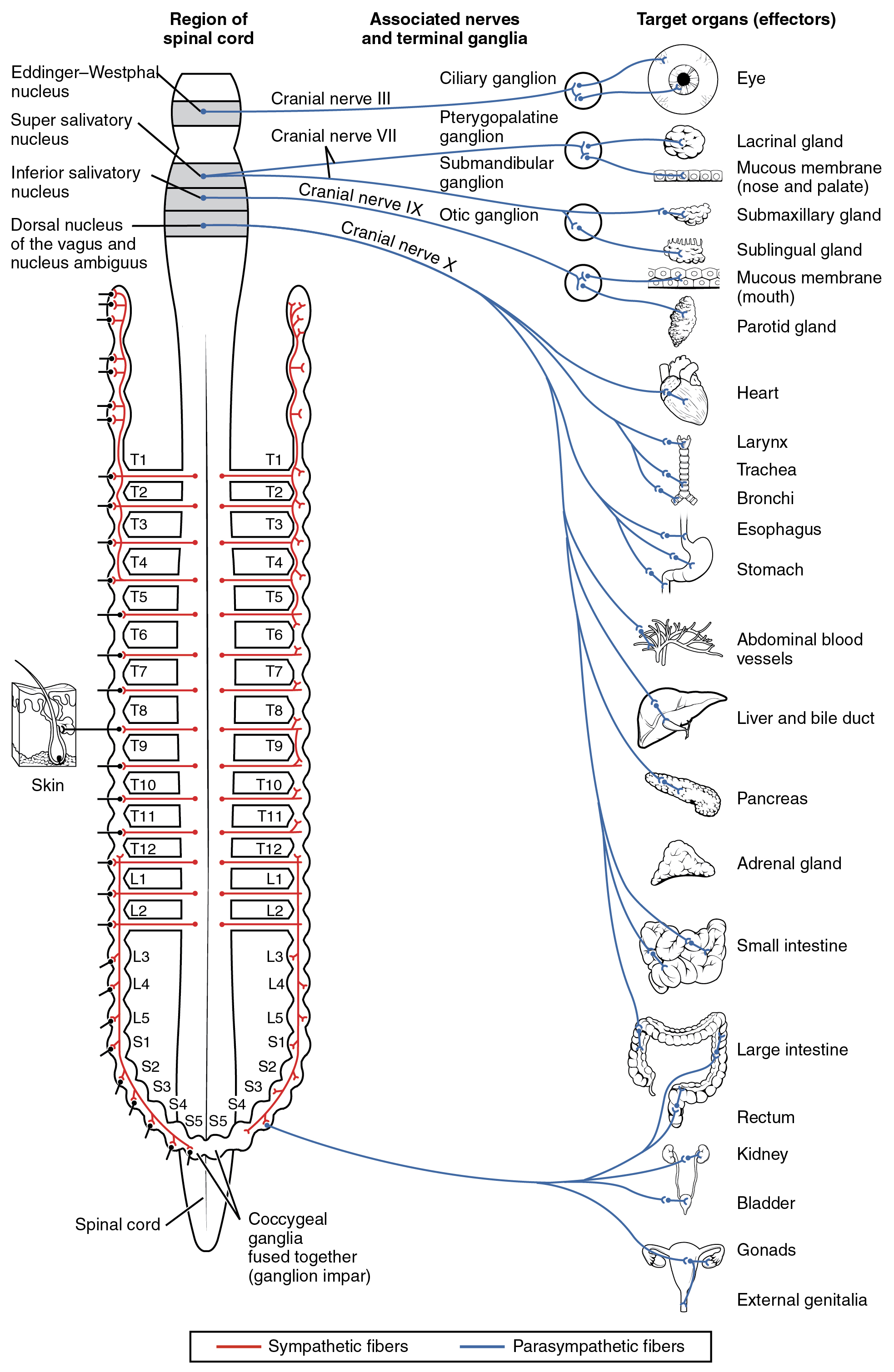
The parasympathetic division of the autonomic nervous arrangement is named because its primal neurons are located on either side of the thoracolumbar region of the spinal cord (para- = "abreast" or "most"). The parasympathetic organisation can besides be referred to every bit the craniosacral system (or outflow) considering the preganglionic neurons are located in nuclei of the encephalon stem and the lateral horn of the sacral spinal cord.
The connections, or "circuits," of the parasympathetic partition are similar to the general layout of the sympathetic division with a few specific differences ([link]). The preganglionic fibers from the cranial region travel in cranial nerves, whereas preganglionic fibers from the sacral region travel in spinal fretfulness. The targets of these fibers are terminal ganglia, which are located virtually—or fifty-fifty within—the target effector. These ganglia are frequently referred to as intramural ganglia when they are found within the walls of the target organ. The postganglionic cobweb projects from the terminal ganglia a short altitude to the target effector, or to the specific target tissue within the organ. Comparing the relative lengths of axons in the parasympathetic arrangement, the preganglionic fibers are long and the postganglionic fibers are short because the ganglia are close to—and sometimes within—the target effectors.
The cranial component of the parasympathetic system is based in particular nuclei of the brain stem. In the midbrain, the Edinger–Westphal nucleus is office of the oculomotor complex, and axons from those neurons travel with the fibers in the oculomotor nerve (cranial nerve 3) that innervate the extraocular muscles. The preganglionic parasympathetic fibers within cranial nervus 3 terminate in the ciliary ganglion, which is located in the posterior orbit. The postganglionic parasympathetic fibers then project to the smooth muscle of the iris to command pupillary size. In the upper medulla, the salivatory nuclei contain neurons with axons that project through the facial and glossopharyngeal nerves to ganglia that control salivary glands. Tear production is influenced by parasympathetic fibers in the facial nervus, which actuate a ganglion, and ultimately the lacrimal (tear) gland. Neurons in the dorsal nucleus of the vagus nervus and the nucleus ambiguus project through the vagus nervus (cranial nerve X) to the terminal ganglia of the thoracic and abdominal cavities. Parasympathetic preganglionic fibers primarily influence the heart, bronchi, and esophagus in the thoracic cavity and the stomach, liver, pancreas, gall bladder, and small intestine of the abdominal crenel. The postganglionic fibers from the ganglia activated by the vagus nervus are often incorporated into the structure of the organ, such as the mesenteric plexus of the digestive tract organs and the intramural ganglia.
Connections of Parasympathetic Sectionalization of the Autonomic Nervous System
Neurons from encephalon-stem nuclei, or from the lateral horn of the sacral spinal cord, project to terminal ganglia near or within the various organs of the body. Axons from these ganglionic neurons then project the short distance to those target effectors.
Chemical Signaling in the Autonomic Nervous System
Where an autonomic neuron connects with a target, there is a synapse. The electrical signal of the action potential causes the release of a signaling molecule, which will bind to receptor proteins on the target cell. Synapses of the autonomic arrangement are classified as either cholinergic, meaning that acetylcholine (ACh) is released, or adrenergic, meaning that norepinephrine is released. The terms cholinergic and adrenergic refer not simply to the signaling molecule that is released but also to the class of receptors that each binds.
The cholinergic system includes two classes of receptor: the nicotinic receptor and the muscarinic receptor. Both receptor types bind to ACh and cause changes in the target cell. The nicotinic receptor is a ligand-gated cation aqueduct and the muscarinic receptor is a Thousand poly peptide–coupled receptor. The receptors are named for, and differentiated by, other molecules that demark to them. Whereas nicotine volition bind to the nicotinic receptor, and muscarine will bind to the muscarinic receptor, there is no cross-reactivity between the receptors. The situation is similar to locks and keys. Imagine 2 locks—one for a classroom and the other for an office—that are opened by two split keys. The classroom key volition not open up the role door and the office primal will not open the classroom door. This is similar to the specificity of nicotine and muscarine for their receptors. Nevertheless, a main key tin open up multiple locks, such as a master primal for the Biology Section that opens both the classroom and the office doors. This is similar to ACh that binds to both types of receptors. The molecules that ascertain these receptors are not crucial—they are simply tools for researchers to use in the laboratory. These molecules are exogenous, significant that they are made exterior of the human body, and so a researcher can use them without any confounding endogenous results (results acquired past the molecules produced in the body).
The adrenergic organization as well has two types of receptors, named the blastoff (α)-adrenergic receptor and beta (β)-adrenergic receptor. Unlike cholinergic receptors, these receptor types are non classified by which drugs tin bind to them. All of them are Yard poly peptide–coupled receptors. There are three types of α-adrenergic receptors, termed α1, αii, and αthree, and there are two types of β-adrenergic receptors, termed β1 and β2. An additional aspect of the adrenergic organisation is that there is a second signaling molecule called epinephrine. The chemical departure between norepinephrine and epinephrine is the addition of a methyl group (CHiii) in epinephrine. The prefix "nor-" actually refers to this chemical departure, in which a methyl group is missing.
The term adrenergic should remind you of the give-and-take adrenaline, which is associated with the fight-or-flight response described at the kickoff of the chapter. Adrenaline and epinephrine are 2 names for the aforementioned molecule. The adrenal gland (in Latin, ad- = "on top of"; renal = "kidney") secretes adrenaline. The ending "-ine" refers to the chemical being derived, or extracted, from the adrenal gland. A similar construction from Greek instead of Latin results in the discussion epinephrine (epi- = "above"; nephr- = "kidney"). In scientific usage, epinephrine is preferred in the U.s., whereas adrenaline is preferred in Nifty Britain, because "adrenalin" was once a registered, proprietary drug proper name in the United States. Though the drug is no longer sold, the convention of referring to this molecule by the ii different names persists. Similarly, norepinephrine and noradrenaline are two names for the same molecule.
Having understood the cholinergic and adrenergic systems, their office in the autonomic system is relatively simple to understand. All preganglionic fibers, both sympathetic and parasympathetic, release ACh. All ganglionic neurons—the targets of these preganglionic fibers—have nicotinic receptors in their cell membranes. The nicotinic receptor is a ligand-gated cation aqueduct that results in depolarization of the postsynaptic membrane. The postganglionic parasympathetic fibers besides release ACh, but the receptors on their targets are muscarinic receptors, which are G protein–coupled receptors and do non exclusively cause depolarization of the postsynaptic membrane. Postganglionic sympathetic fibers release norepinephrine, except for fibers that project to sweat glands and to blood vessels associated with skeletal muscles, which release ACh ([link]).
| Autonomic System Signaling Molecules | ||
|---|---|---|
| Sympathetic | Parasympathetic | |
| Preganglionic | Acetylcholine → nicotinic receptor | Acetylcholine → nicotinic receptor |
| Postganglionic | Norepinephrine → α- or β-adrenergic receptors Acetylcholine → muscarinic receptor (associated with sweat glands and the blood vessels associated with skeletal muscles only | Acetylcholine → muscarinic receptor |
Signaling molecules can belong to 2 broad groups. Neurotransmitters are released at synapses, whereas hormones are released into the bloodstream. These are simplistic definitions, merely they tin can aid to clarify this point. Acetylcholine tin be considered a neurotransmitter because it is released by axons at synapses. The adrenergic system, nonetheless, presents a challenge. Postganglionic sympathetic fibers release norepinephrine, which can be considered a neurotransmitter. But the adrenal medulla releases epinephrine and norepinephrine into circulation, so they should be considered hormones.
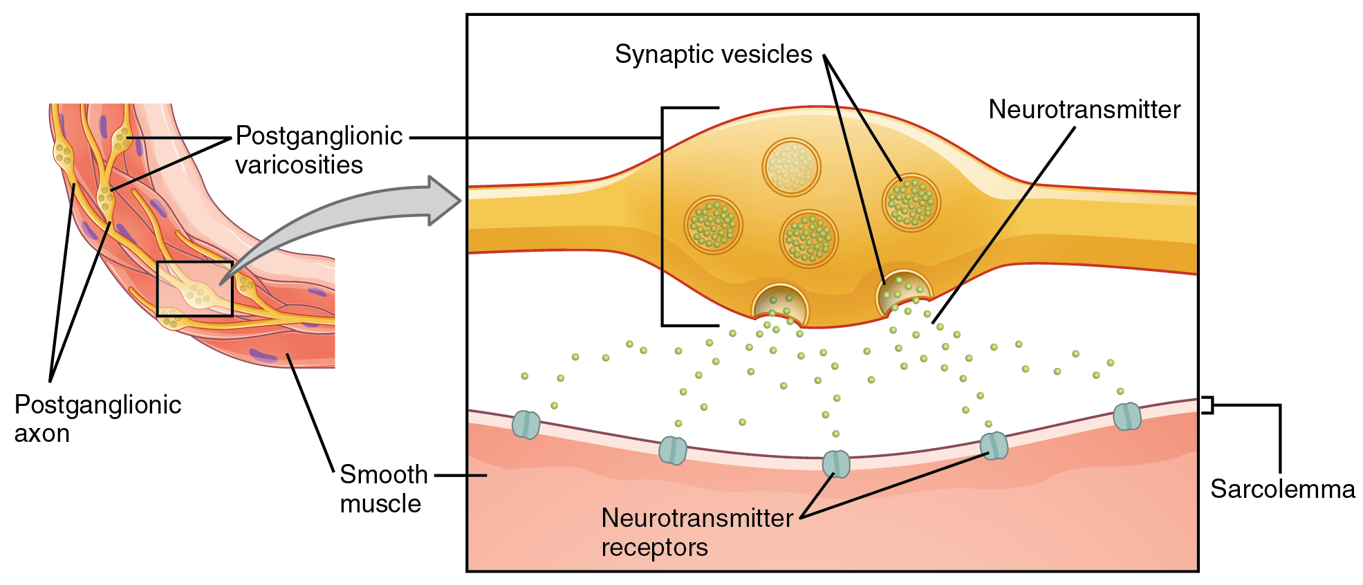
What are referred to hither as synapses may non fit the strictest definition of synapse. Some sources volition refer to the connection between a postganglionic cobweb and a target effector as neuroeffector junctions; neurotransmitters, as defined to a higher place, would exist called neuromodulators. The structure of postganglionic connections are not the typical synaptic terminate bulb that is found at the neuromuscular junction, but rather are chains of swellings along the length of a postganglionic fiber called a varicosity ([link]).
Autonomic Varicosities
The connection between autonomic fibers and target effectors is not the same as the typical synapse, such as the neuromuscular junction. Instead of a synaptic stop seedling, a neurotransmitter is released from swellings along the length of a fiber that makes an extended network of connections in the target effector.
Everyday Connections
Fight or Flight? What About Fear and Freeze?
The original usage of the epithet "fight or flying" comes from a scientist named Walter Cannon who worked at Harvard in 1915. The concept of homeostasis and the functioning of the sympathetic system had been introduced in France in the previous century. Cannon expanded the idea, and introduced the idea that an beast responds to a threat past preparing to stand and fight or run away. The nature of this response was thoroughly explained in a volume on the physiology of pain, hunger, fear, and rage.
When students larn about the sympathetic arrangement and the fight-or-flight response, they often end and wonder nearly other responses. If yous were faced with a lioness running toward you as pictured at the offset of this affiliate, would you run or would you stand up your footing? Some people would say that they would freeze and not know what to do. Then isn't in that location really more to what the autonomic system does than fight, flight, rest, or digest. What well-nigh fright and paralysis in the confront of a threat?
The common epithet of "fight or flight" is beingness enlarged to be "fight, flight, or fright" or fifty-fifty "fight, flying, fright, or freeze." Cannon's original contribution was a catchy phrase to limited some of what the nervous organization does in response to a threat, but it is incomplete. The sympathetic system is responsible for the physiological responses to emotional states. The name "sympathetic" tin be said to hateful that (sym- = "together"; -pathos = "pain," "suffering," or "emotion").

Sentinel this video to learn more virtually the nervous system. As described in this video, the nervous system has a style to deal with threats and stress that is separate from the conscious control of the somatic nervous organization. The system comes from a fourth dimension when threats were virtually survival, but in the modern age, these responses become part of stress and anxiety. This video describes how the autonomic organization is but part of the response to threats, or stressors. What other organ system gets involved, and what role of the brain coordinates the 2 systems for the entire response, including epinephrine (adrenaline) and cortisol?
Chapter Review
The principal responsibilities of the autonomic nervous organisation are to regulate homeostatic mechanisms in the body, which is also part of what the endocrine system does. The key to understanding the autonomic system is to explore the response pathways—the output of the nervous system. The style we respond to the world effectually u.s.a., to manage the internal environs on the ground of the external surround, is divided between two parts of the autonomic nervous system. The sympathetic partitioning responds to threats and produces a readiness to confront the threat or to run away: the fight-or-flight response. The parasympathetic segmentation plays the reverse role. When the external environment does not present any firsthand danger, a restful mode descends on the body, and the digestive system is more active.
The sympathetic output of the nervous system originates out of the lateral horn of the thoracolumbar spinal cord. An axon from one of these key neurons projects past way of the ventral spinal nervus root and spinal nerve to a sympathetic ganglion, either in the sympathetic chain ganglia or one of the collateral locations, where it synapses on a ganglionic neuron. These preganglionic fibers release ACh, which excites the ganglionic neuron through the nicotinic receptor. The axon from the ganglionic neuron—the postganglionic fiber—then projects to a target effector where it volition release norepinephrine to demark to an adrenergic receptor, causing a alter in the physiology of that organ in keeping with the broad, divergent sympathetic response. The postganglionic connections to sweat glands in the pare and claret vessels supplying skeletal muscle are, even so, exceptions; those fibers release ACh onto muscarinic receptors. The sympathetic system has a specialized preganglionic connection to the adrenal medulla that causes epinephrine and norepinephrine to be released into the bloodstream rather than exciting a neuron that contacts an organ directly. This hormonal component ways that the sympathetic chemical point tin spread throughout the trunk very rapidly and affect many organ systems at once.
The parasympathetic output is based in the brain stem and sacral spinal cord. Neurons from detail nuclei in the brain stem or from the lateral horn of the sacral spinal string (preganglionic neurons) project to last (intramural) ganglia located close to or within the wall of target effectors. These preganglionic fibers as well release ACh onto nicotinic receptors to excite the ganglionic neurons. The postganglionic fibers then contact the target tissues within the organ to release ACh, which binds to muscarinic receptors to induce rest-and-assimilate responses.
Signaling molecules utilized past the autonomic nervous system are released from axons and can be considered as either neurotransmitters (when they directly interact with the effector) or as hormones (when they are released into the bloodstream). The aforementioned molecule, such as norepinephrine, could be considered either a neurotransmitter or a hormone on the basis of whether information technology is released from a postganglionic sympathetic axon or from the adrenal gland. The synapses in the autonomic system are not ever the typical type of connection beginning described in the neuromuscular junction. Instead of having synaptic end bulbs at the very end of an axonal fiber, they may accept swellings—called varicosities—along the length of a fiber then that information technology makes a network of connections inside the target tissue.
Interactive Link Questions
Watch this video to learn more most adrenaline and the fight-or-flight response. When someone is said to take a blitz of adrenaline, the image of bungee jumpers or skydivers usually comes to mind. But adrenaline, also known every bit epinephrine, is an important chemic in analogous the body's fight-or-flight response. In this video, you look inside the physiology of the fight-or-flight response, as envisioned for a firefighter. His torso'due south reaction is the result of the sympathetic division of the autonomic nervous system causing organisation-broad changes as it prepares for extreme responses. What two changes does adrenaline bring nearly to help the skeletal muscle response?
The heart charge per unit increases to send more claret to the muscles, and the liver releases stored glucose to fuel the muscles.
Sentry this video to learn more than about the nervous system. As described in this video, the nervous system has a fashion to deal with threats and stress that is separate from the witting control of the somatic nervous system. The system comes from a time when threats were most survival, only in the modernistic historic period, these responses become part of stress and anxiety. This video describes how the autonomic arrangement is only part of the response to threats, or stressors. What other organ system gets involved, and what part of the brain coordinates the two systems for the entire response, including epinephrine (adrenaline) and cortisol?
The endocrine system is as well responsible for responses to stress in our lives. The hypothalamus coordinates the autonomic response through projections into the spinal cord and through influence over the pituitary gland, the constructive heart of the endocrine system.
Review Questions
Which of these physiological changes would not be considered part of the sympathetic fight-or-flight response?
- increased eye rate
- increased sweating
- dilated pupils
- increased stomach motility
D
Which type of fiber could be considered the longest?
- preganglionic parasympathetic
- preganglionic sympathetic
- postganglionic parasympathetic
- postganglionic sympathetic
A
Which signaling molecule is most likely responsible for an increment in digestive activeness?
- epinephrine
- norepinephrine
- acetylcholine
- adrenaline
C
Which of these cranial nerves contains preganglionic parasympathetic fibers?
- optic, CN 2
- facial, CN VII
- trigeminal, CN V
- hypoglossal, CN XII
B
Which of the post-obit is not a target of a sympathetic preganglionic cobweb?
- intermural ganglion
- collateral ganglion
- adrenal gland
- chain ganglion
A
Critical Thinking Questions
In the context of a lioness hunting on the savannah, why would the sympathetic system not activate the digestive system?
Whereas energy is needed for running abroad from the threat, blood needs to be sent to the skeletal muscles for oxygen supply. The additional fuel, in the form of carbohydrates, probably wouldn't improve the ability to escape the threat as much every bit the diversion of oxygen-rich claret would hinder it.
A target effector, such as the heart, receives input from the sympathetic and parasympathetic systems. What is the actual divergence between the sympathetic and parasympathetic divisions at the level of those connections (i.e., at the synapse)?
The postganglionic sympathetic cobweb releases norepinephrine, whereas the postganglionic parasympathetic fiber releases acetylcholine. Specific locations in the eye have adrenergic receptors and muscarinic receptors. Which receptors are bound is the signal that determines how the centre responds.
Glossary
- blastoff (α)-adrenergic receptor
- one of the receptors to which epinephrine and norepinephrine bind, which comes in three subtypes: α1, αtwo, and α3
- acetylcholine (ACh)
- neurotransmitter that binds at a motor end-plate to trigger depolarization
- adrenal medulla
- interior portion of the adrenal (or suprarenal) gland that releases epinephrine and norepinephrine into the bloodstream as hormones
- adrenergic
- synapse where norepinephrine is released, which binds to α- or β-adrenergic receptors
- beta (β)-adrenergic receptor
- one of the receptors to which epinephrine and norepinephrine bind, which comes in two subtypes: β1 and βii
- celiac ganglion
- one of the collateral ganglia of the sympathetic system that projects to the digestive arrangement
- central neuron
- specifically referring to the cell torso of a neuron in the autonomic system that is located in the cardinal nervous system, specifically the lateral horn of the spinal cord or a brain stem nucleus
- cholinergic
- synapse at which acetylcholine is released and binds to the nicotinic or muscarinic receptor
- chromaffin cells
- neuroendocrine cells of the adrenal medulla that release epinephrine and norepinephrine into the bloodstream as part of sympathetic arrangement activity
- ciliary ganglion
- one of the last ganglia of the parasympathetic system, located in the posterior orbit, axons from which projection to the iris
- collateral ganglia
- ganglia exterior of the sympathetic concatenation that are targets of sympathetic preganglionic fibers, which are the celiac, inferior mesenteric, and superior mesenteric ganglia
- craniosacral organization
- alternating name for the parasympathetic division of the autonomic nervous arrangement that is based on the anatomical location of fundamental neurons in brain-stalk nuclei and the lateral horn of the sacral spinal cord; also referred to as craniosacral outflow
- dorsal nucleus of the vagus nerve
- location of parasympathetic neurons that project through the vagus nerve to terminal ganglia in the thoracic and intestinal cavities
- Eddinger–Westphal nucleus
- location of parasympathetic neurons that projection to the ciliary ganglion
- endogenous
- describes substance made in the man body
- epinephrine
- signaling molecule released from the adrenal medulla into the bloodstream as role of the sympathetic response
- exogenous
- describes substance made exterior of the human being body
- fight-or-flight response
- set of responses induced by sympathetic action that pb to either fleeing a threat or standing upward to it, which in the modern world is often associated with anxious feelings
- 1000 protein–coupled receptor
- membrane protein complex that consists of a receptor protein that binds to a signaling molecule—a G protein—that is activated past that binding and in turn activates an effector protein (enzyme) that creates a second-messenger molecule in the cytoplasm of the target cell
- ganglionic neuron
- specifically refers to the cell body of a neuron in the autonomic organisation that is located in a ganglion
- gray rami communicantes
- (singular = ramus communicans) unmyelinated structures that provide a brusque connexion from a sympathetic chain ganglion to the spinal nerve that contains the postganglionic sympathetic fiber
- greater splanchnic nerve
- nerve that contains fibers of the primal sympathetic neurons that do not synapse in the chain ganglia but project onto the celiac ganglion
- inferior mesenteric ganglion
- one of the collateral ganglia of the sympathetic system that projects to the digestive system
- intramural ganglia
- final ganglia of the parasympathetic system that are institute within the walls of the target effector
- lesser splanchnic nervus
- nerve that contains fibers of the central sympathetic neurons that do not synapse in the chain ganglia only project onto the junior mesenteric ganglion
- ligand-gated cation aqueduct
- ion channel, such as the nicotinic receptor, that is specific to positively charged ions and opens when a molecule such as a neurotransmitter binds to it
- mesenteric plexus
- nervous tissue within the wall of the digestive tract that contains neurons that are the targets of autonomic preganglionic fibers and that project to the polish muscle and glandular tissues in the digestive organ
- muscarinic receptor
- type of acetylcholine receptor protein that is characterized by also bounden to muscarine and is a metabotropic receptor
- nicotinic receptor
- type of acetylcholine receptor protein that is characterized by besides binding to nicotine and is an ionotropic receptor
- norepinephrine
- signaling molecule released every bit a neurotransmitter by almost postganglionic sympathetic fibers every bit part of the sympathetic response, or as a hormone into the bloodstream from the adrenal medulla
- nucleus ambiguus
- brain-stem nucleus that contains neurons that project through the vagus nerve to last ganglia in the thoracic crenel; specifically associated with the heart
- parasympathetic division
- division of the autonomic nervous system responsible for restful and digestive functions
- paravertebral ganglia
- autonomic ganglia superior to the sympathetic chain ganglia
- postganglionic cobweb
- axon from a ganglionic neuron in the autonomic nervous organisation that projects to and synapses with the target effector; sometimes referred to as a postganglionic neuron
- preganglionic fiber
- axon from a fundamental neuron in the autonomic nervous system that projects to and synapses with a ganglionic neuron; sometimes referred to as a preganglionic neuron
- prevertebral ganglia
- autonomic ganglia that are anterior to the vertebral column and functionally related to the sympathetic chain ganglia
- rest and digest
- set of functions associated with the parasympathetic system that lead to restful actions and digestion
- superior cervical ganglion
- one of the paravertebral ganglia of the sympathetic system that projects to the caput
- superior mesenteric ganglion
- ane of the collateral ganglia of the sympathetic system that projects to the digestive system
- sympathetic chain ganglia
- serial of ganglia side by side to the vertebral column that receive input from central sympathetic neurons
- sympathetic division
- partitioning of the autonomic nervous system associated with the fight-or-flight response
- target effector
- organ, tissue, or gland that will respond to the control of an autonomic or somatic or endocrine signal
- terminal ganglia
- ganglia of the parasympathetic sectionalisation of the autonomic system, which are located near or inside the target effector, the latter also known every bit intramural ganglia
- thoracolumbar system
- alternate name for the sympathetic division of the autonomic nervous arrangement that is based on the anatomical location of fundamental neurons in the lateral horn of the thoracic and upper lumbar spinal cord
- varicosity
- construction of some autonomic connections that is not a typical synaptic end bulb, only a cord of swellings along the length of a fiber that makes a network of connections with the target effector
- white rami communicantes
- (atypical = ramus communicans) myelinated structures that provide a short connection from a sympathetic chain ganglion to the spinal nerve that contains the preganglionic sympathetic fiber

Which Of The Following Does The Autonomic Nervous System Most Directly Control?,
Source: https://pressbooks-dev.oer.hawaii.edu/anatomyandphysiology/chapter/divisions-of-the-autonomic-nervous-system/
Posted by: lapplefuld.blogspot.com
0 Response to "Which Of The Following Does The Autonomic Nervous System Most Directly Control?"
Post a Comment